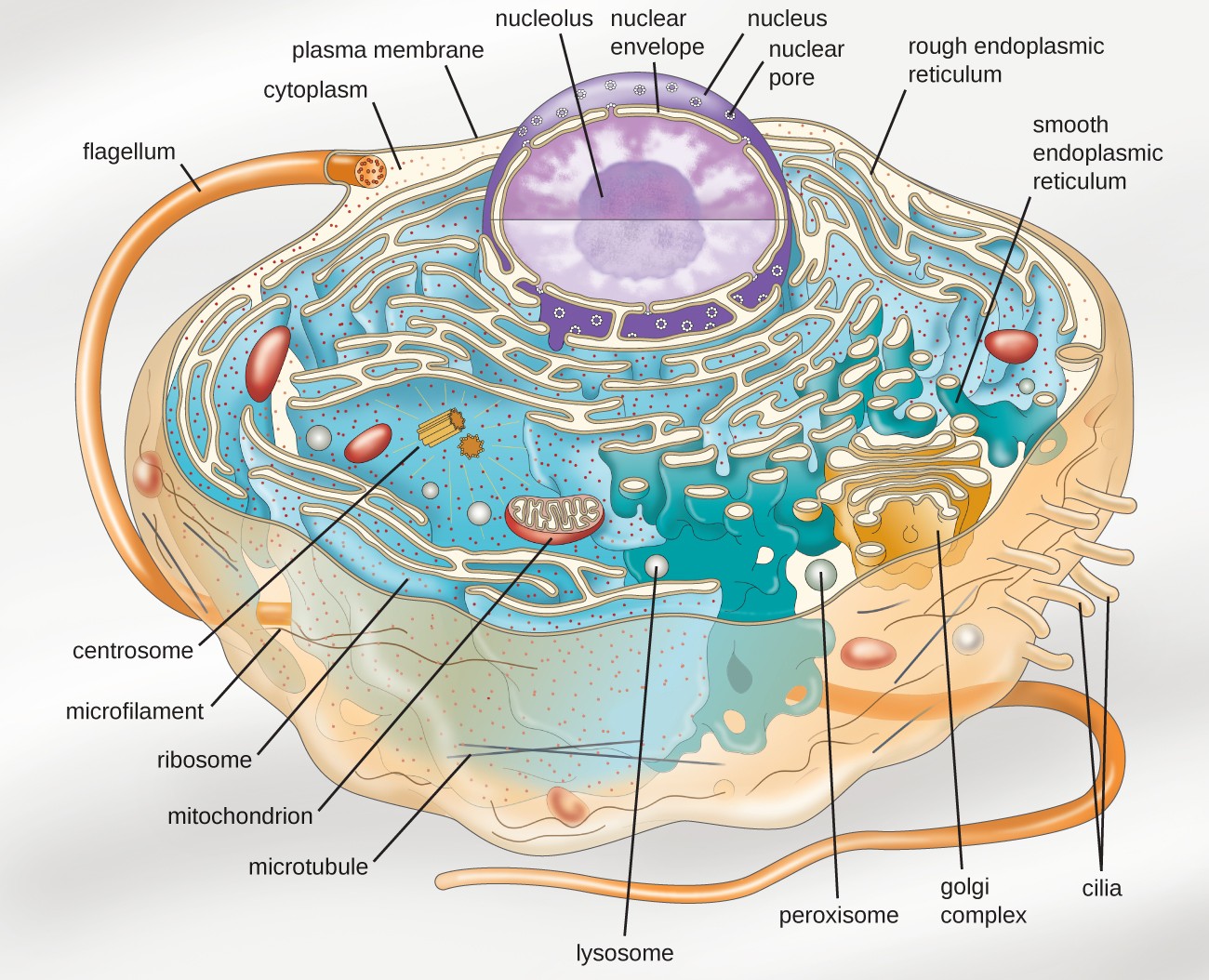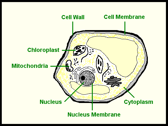

May require additional steps for sample preparation and staining May require optimization for different experimental conditionsĪllows for localization and tracking of specific cellular components May require optimization for different cell types and applicationsĬompatible with various imaging techniques Advantages and disadvantages of fluorescent dyesĬan be used for both fixed and live cellsĬan be multiplexed for simultaneous labelling of multiple targets GFP has allowed researchers to track biological processes in real-time, providing insights into the behaviour of cells that were previously impossible or really hard to observe. Originally discovered in the bioluminescent jellyfish Aequorea Victoria, GFP has since been isolated and extensively studied. Green Fluorescent Protein, or GFP for short, is a protein that has revolutionized the field of molecular biology. Synthetic dyes can be used to label biomolecules such as proteins, antibodies, peptides, nucleic acid, yeast and bacteria. The fluorophore then emits light to liberate the extra energy it absorbed.įluorophores can be designed to bind to specific cell structures or components, and to absorb or emit light at a specific wavelength, allowing different types of fluorescent dyes to be combined in the same staining.įluorescent dyes can occur naturally in the living world, like the case of the Green Fluorescent Protein (GFP), or can be made synthetically. In other words, light reaches the fluorophore, which absorbs it and increases its energy. A fluorophore is a molecule that can emit light after getting excited by light energy. We will have a look at the more advanced methods of cell labelling in this article, but you can check out our Gram-Staining article to learn more about this method! Fluorescent Dyes as Cell Labelling Methodsįluorescent dyes are biological molecules composed of at least one fluorophore. Therefore, when wanting to track a cellular process in real-time through the use of dyes, it's important to know if the process will kill the cells or keep them alive. Depending on the research question and the type of technique, cells might be "fixed" and killed during the staining process. These all give us ways to see the structures of a cell more easily. These include some of the most basic laboratory techniques like Gram staining, or more complex techniques like using fluorescent dyes, immunolabeling or fluorescent fusion proteins. Cell Labelling MethodsĬells can be labelled in multiple ways. Therefore, electron micrographs of cells are detailed images of the cells in which organelles can be identified and labelled. Electron microscopes are essential for observing smaller organelles such as ribosomes, vesicles, granules, and filaments that cannot be seen directly with light microscopy. This allows them to capture clearer images from the cells with much higher definition. Other organelles are smaller than the resolution of light microscopes, so they appear blurry and not well-defined under light microscopes.Įlectron microscopes, on the other hand, are much stronger in magnification and resolution. The capacity of a microscope to generate a picture of an item at a scale bigger than its true size is referred to as magnification.įor instance, organelles visible under a light microscope include the nucleus, cytoplasm, cell membrane, chloroplasts, and the cell wall.

In different words, the resolution is the smallest distance between two different points of a specimen that may still be viewed as independent entities when seen under the microscope.


The term ' resolution' or ' resolving power' in microscopy refers to a microscope's capacity to see detail. Non Specific Defences of the Human Body.


 0 kommentar(er)
